In biomedicine, accurate and efficient observation of pathologic slices is crucial in cell morphology detection, pathologic analysis, and disease diagnosis, which acts as the bridge between fundamental research and clinical applications. On the one hand, pathologic slices are usually stained for specific recognition, given the fact that humans are sensitive to color information and capable of classification according to color. On the other hand, digital pathology that uses digital cameras to collect stained pathologic slices improves imaging efficiency compared with the naked eyes and reduces oversight and double counting. However, a tradeoff between high resolution (HR) and wide field of view (FOV) exists in digital pathology, resulting in artifacts of scanning and stitching.
Fourier ptychographic microscopy (FPM), invented in 2013 by Zheng and Yang et al., is a promising computational imaging technique that eliminates these artifacts in digital pathology and provides a high throughput, sharing its root with optical phase retrieval and synthetic aperture radar. Given its flexible setup, performance without mechanical scanning, and interferometric measurements, FPM has successful applications in digital pathology and whole slide imaging systems.
Currently, the conventional full-color digital pathology based on FPM is still time-consuming because of the repeated experiments with tri-wavelengths. Inspired by color matching, Prof. PAN An, Prof. YAO Baoli, and Prof. MA Caiwen at Xi'an Institute of Optics and Precision Mechanics (XIOPM), Chinese Academy of Sciences (CAS) reported a colorization method via color transfer termed CFPM. The reconstruction time is significantly reduced by 2/3 with the sacrifice of precision of only 0.4%, which marks a great leap for the efficiency of FPM colorization compared with traditional methods. Besides, CFPM is easy to operate and promote without requirements in terms of overlapping rate, sampling rate or training data set. And CFPM can be regarded as an “unsupervised transfer learning” based on physical models without iterative optimization in contrast to traditional transfer learning. This may provide new ideas for related work in the future.
The study was published inScience China-Physics Mechanics & Astronomy (IF=5.122) on July 27th and was selected as the cover story of issue 11.
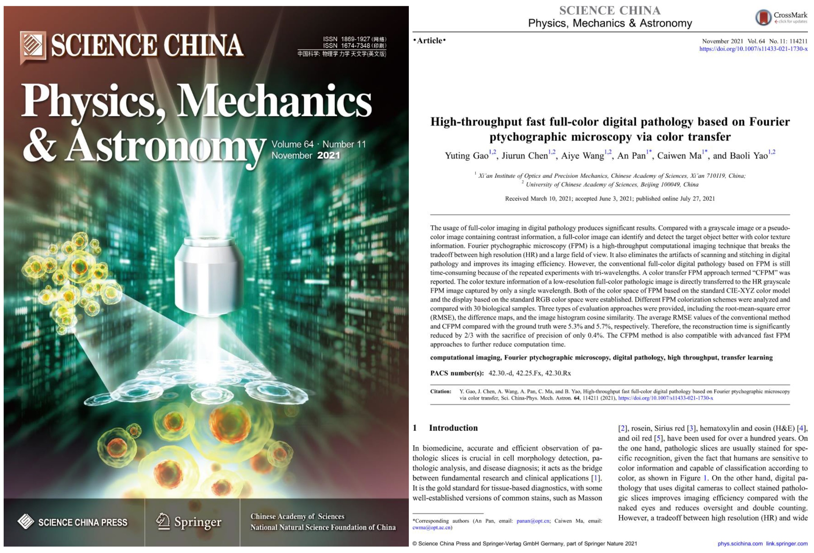
Figure 1. cover and the first page of the paper
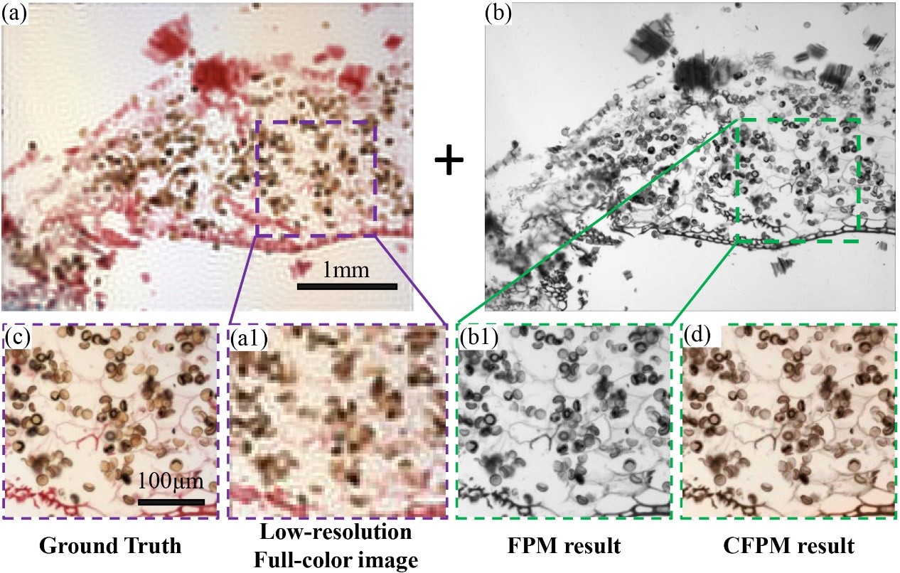
 Figure 2 Results of stained resting sporangia. (a, a1) LR donor image with the entire FOV of a 4× / 0.1NA objective and its close-up. (b, b1) FPM recovery image under green channel (515.0 nm) and its close-up. (c) Ground truth captured by a 10× / 0.3NA objective. (d) Staining results via CFPM.
There are two technical difficulties: one is how to ensure the authenticity of color and the correctness during displaying process; The other one is how to ensure the accuracy of color transfer while improving efficiency. Therefore, the mapping relationship between CIE-XYZ color space and the display of different color spaces is established; Different color transfer schemes are compared and the result shows that it is the best option to use low-resolution color images with the same field of view as donor images. It is also proved that low-resolution color images used for color transfer have enough color texture information.
Speaking of the future application, Prof. PAN An, the corresponding author of the paper, said: “By transferring low-resolution true color texture information of optical microscopes to electronic microscopes, this method also enlightens us that we may dye true color for black-and-white images of an electronic microscope”.
 Figure 3. Examples of future application


