Malignant tumor (cancer) is a new organism formed by mutation of normal cells in vivo. It can grow continuously and escape the elimination of the immune system, which become the leading cause of death for its high incidence and mortality. Therefore, Early diagnosis and treatment are essential for the inhibition of tumor growth.
In contrast to conventional means, bioimaging has the potential to accurately locate and diagnose tumors at an early stage by the methods of characterization, visualization, and quantification of physiological processes at the cellular and molecular levels. However, due to the low resolution and dangerous ionizing radiation of CT, PET and SPECT imaging, as well as the high time-consuming of PAI and MRI imaging, their application in disease treatment is limited. Are there efficient methods for tumor diagnosis?
A research team led by Prof. Dr. PENG Bo from Xi'an Institute of Optics and Precision Mechanics (XIOPM) of the Chinese Academy of Sciences (CAS) proposed a biocompatible, nontoxic, near-infrared fluorescent tumor targeting nanoparticles (NPs) KHLF(K5HoLi2F10) for the early diagnosis of tumor. The results were published in JOURNAL OF LUMINESCENCE.
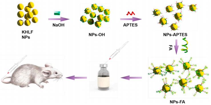 Schematic illustration of the preparation of FA-NPs and its bio-application. (Image by XIOPM)
The preparation of the KHLF NPs is according to the atypical hydrothermal method:
CH3CH2OK, Ho(NO3)3 ● 6H2O, and LiNO3 were respectively prepared into aqueous solutions of a certain concentration by being dissolved in deionized water. After mixing them, PEG was added to the mixture and stirred evenly.
“We modified APTES on the novel NPs doped with Ho3+ by a step-by-step method and covalently linked folic acid (FA) to the surface to endow it with the water-solubility and tumor targeting”. The results of FTIR and zeta potential confirmed the modification process.
From the subsequent fluorescence imaging, the modification of FA did not significantly weaken the fluorescence intensity of NPs. Therefore, the biodistribution imaging, in-vivo imaging, in-vitro imaging of various organs, and tumor targeting imaging experiments are carried out. The results show that this nanoparticle can accurately accumulate on HeLa tumor and a generate strong fluorescence signal.
The proposed fluorescence imaging based on nanoparticle has great potential as an efficient biocompatible fluorescent contrast agent for tumor diagnosis.