As an increasingly attractive technique, light-sheet fluorescence microscopy (LSFM) has made considerable progress over the past decade in the fields of developmental biology, whole-brain imaging, single molecules detection, etc. the LSFM inherently has its unique advantages such as a good optical sectioning capability and a good signal-to-noise ratio (SNR).Besides, the wide-field imaging mode permits LSFM an intrinsic fast imaging technique.
However, when a high axial resolution or optical sectioning capability is required, the field of view (FOV) is limited. This is because that the FOV depends on the size of the light-sheet, especially the length of the light-sheet limited by the Rayleigh length of the excitation beam. Thus, some compromises still need to be made among the properties of the light-sheet, including the axial resolution/ optical sectioning capability and the FOV.

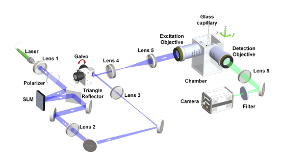
The schematic diagram of the LSFM system (Image by XIOPM)
To breakthrough this limitation, Gao et al. presented a method by tiling a relatively small light-sheet sequentially to different positions within the image plane. But this method is too sophisticated for that, for each tiling position within the image plane, a corresponding image should be taken and stitched together to obtain a large FOV. Thus, the imaging speed has to be traded as a substitute for both a large FOV and a high axial resolution.
Now a research team led by Prof. Dr. YAO Baoli from Xi'an Institute of Optics and Precision Mechanics (XIOPM) of the Chinese Academy of Sciences (CAS) propose an alternative FOV extension method by scanning the multiple focused shifted Gaussian beams array (MGBA) along the direction perpendicular to the propagation axis. The result was published in OPTTICS EXPRESS.
In this method, the positions of the beam waists of the multiple Gaussian beam arrays are shifted in both axial and lateral directions in an optimized arranged pattern, and then scanned along the direction perpendicular to the propagation axis to form an extended FOV of light-sheet. Complementary beam subtraction method is also adopted to further improve axial resolution.

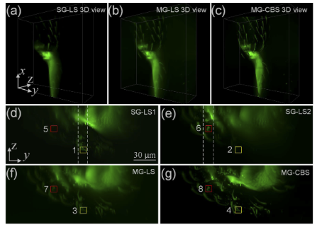
Imaging of the hind leg of flea beetle (Image by XIOPM)
The imaging performance was proved to have a large FOV without sacrificing the axial resolution or optical sectioning capability by imaging fluorescent beads and the hind leg of flea beetle.
Moreover, the interval and the distribution of the beams in the MGBA can be changed in the algorithm to generate different sizes of FOV. Such ability and flexibility make it suitable to apply to the ROI imaging, including discrete ROI in the same FOV, which can improve the energy efficiency or reduce the unnecessary photobleaching and phototoxicity to the specimen.


