Fluorescence imaging has been acting as a mainstream observation technique in the clinic diagnosis, but the fluorescence imaging approaches suffer from the scattering and absorption of the light by tissues, especially under the situation of visible‐light‐emitting fluorescence probes are used.
To overcome the problem, a research team led by Prof. Dr. PENG Bo from Xi'an Institute of Optics and Precision Mechanics (XIOPM) of the Chinese Academy of Sciences (CAS) developed novel near‐infrared (NIR) region fluorescent PEG400‐K5HoLi2F10 (KHLF) nanoparticles (NPs) with exciting photothermal performance by a simple hydrothermal method. The results were published in JOURNAL OF BIOMEDICAL MATERIALS RESEARCH.
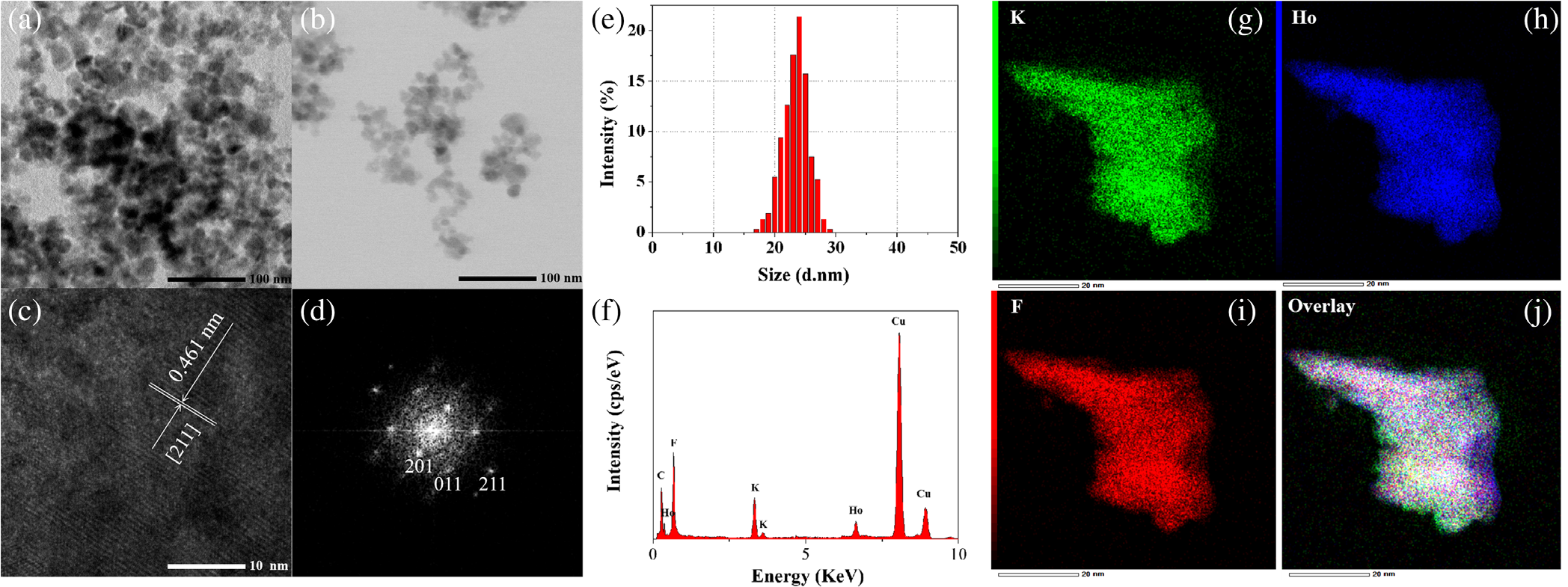 Transmission electron microscope (TEM) images of unmodified K5HoLi2F10 (KHLF) nanoparticles (NPs) (a) and the ones with modification by PEG400 (b). High-resolution TEM (HR-TEM) images (c) and selected area electron diffraction patterns (d) of the sample KHLF. The dynamic light scattering (DLS) spectrum of sample KHLF (e). Energy dispersive X-ray spectrometer (EDS) spectrum (f) and the element distribution of KHLF (g–j). (Image by XIOPM)
Compared with conventional NaYF4 NPs, the KHLF as a novel doping matrix exhibits superior optical properties, The NPs exhibit high near‐infrared (NIR) region fluorescence emission efficiency and superior photostability.
Furthermore, the superior performance of this nanoprobe for in vivo imaging was verified by imaging living mice. The fluorescence image shows a significant SBR of 14:1 and low biotoxicity, rapid metabolism, and biodistribution, indicating the KHLF NPs have a great potential for noninvasive imaging and diagnosis of tumor in vivo.
This KHLF‐based nanoplatform also combines the fluorescence imaging and photothermal imaging which demonstrates the advantages of collaborative diagnosis than the single diagnostic model in vivo.