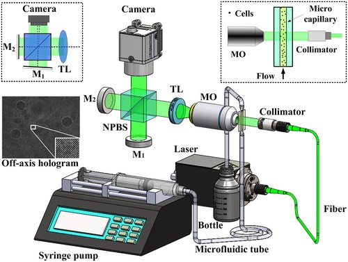
Baoli Yao’s research team combined Michelson‐interferometer‐based off‐axis digital holographic microscopy (DHM) with a common flow cytometry (FCM) arrangement. Utilizing object recognition procedures and holographic autofocusing during the numerical reconstruction of the acquired off‐axis holograms, sharply focused quantitative phase images of suspended cells in flow were retrieved without labeling, from which biophysical cellular features of distinct cells, such as cell radius, refractive index and dry mass, can be subsequently retrieved in an automated manner. The performance of the proposed concept was first characterized by investigations on microspheres that were utilized as test standards. Then, they analyzed two types of pancreatic tumor cells with different morphology to further verify the applicability of the proposed method for quantitative live cell imaging. The retrieved biophysical datasets from cells in flow are found in good agreement with results from comparative investigations with previously developed DHM methods under static conditions, which demonstrates the effectiveness and reliability of our approach. Our results contribute to the establishment of DHM in imaging FCM and prospect to broaden the application spectrum of FCM by providing complementary quantitative imaging as well as additional biophysical cell parameters which are not accessible in current high‐throughput FCM measurements.
(Original research article " J. Biophotonics. 2019;12:e201900085 https://doi.org/10.1002/jbio.201900085)