Optics Express, a famous optical journal under the Optical Society of America, published three research papers by the Yao Baoli Research Group under the National Key Laboratory for Transient Optics and Photon Technology of our institute on Feb. 18, 2019, which is the first event in the history of our institute.
Fig.1.Optics Express published three research papers by the Yao Baoli Research Group under the National Key Laboratory for Transient Optics and Photon Technology of our institute
The first paper “Large-scale 3D Imaging of Insects with Natural Color” is of how to realize large-scale 3D high-resolution quantitative imaging of insects with natural color. As we know, after the evolution in hundreds of millions of years, biostructure becomes very complicated and exquisite, and bears diversified functions and fascinating scenes. The observation and form analysis of biostructure by different scale, dimension and position prepares the most direct evidence for scientific research results, and plays an essential role in numerous disciplines and fields. Currently, high-resolution 3D imaging technology has been widely used in biology, and fueled new progress in relevant studies. However, there are some deficiencies in existing technologies and research instruments. For example, when carrying out 3D imaging of large samples, the data volume is high and the process is time-consuming; it is hard to satisfy the needs of high resolution and large imaging view field at the same time, and to acquire natural color of the sample as well as others. Therefore, it becomes an urgent need of insect taxology scientists and relevant research fields to identify a kind of equipment able to realize quick 3D imaging of insects and acquire their high-resolution shape and appearance information and color information.
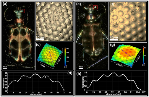
Fig. 2. 3D Imaging Results of Two Kinds of Chinese Tiger Beetles. (a) The highest-value projection of Tiger Beetle 1 (4X, NA0.2), with the 3D imaging volume of about 18.7 x 9.4 x 7.0 mm3. (b) The highest-value projection results from the imaging of the area in the red block in Fig.(a) with 20X, NA0.45 objective. (c) 3D shape and appearance information in Fig. (b). (d) The compound eye’s 3D profile curve that the blue curve in Fig. (c) goes through. (e) The highest-value projection of Tiger Beetle 2 (4X, NA0.2),with the 3D imaging volume of about 19.5 x 8.3 x 6.6 mm3. (f)The highest-value projection results from the imaging of area in the red block in Fig. (e) with 20X, NA0.45 objective. (g) 3D shape and appearance information in Fig.(f). (h) The compound eye’s 3D profile curve that the blue curve in Fig. (g) goes through.
The second paper “Real-time Optical Manipulation of Particles through Turbid Media” mainly realizes real-time optical micro manipulation of particles after penetrating scattering medium. It is known that half of the Nobel Prize in Physics 2018 was granted to Arthur Ashkin, the inventor of optical tweezers, with which capturing and manipulating particles by laser is carried out in transparent and scattering-medium-free environment. When any scattering medium exists in the optical system, imaging object will be hard to present clearly on the image surface, and laser is hard to focus on a focal point. At present, there are multiple methods to overcome influences of scattering, of which the most common is that regulating the optical field after scattering medium with optical field regulating device and corresponding optimization algorithm. Genetic algorithm features quick convergence and strong noise resistance, so it has been widely used in focusing and imaging of optical field after scattering medium. However, there are still certain problems with genetic algorithm in actual application. For example, with optimization going, its convergence rate will become slow gradually; noise makes high influences on the final focusing results; and the optimization results are limited by the probe’s dynamic range, etc. In recent years, with relevant technologies becoming mature, some researchers have combined wave-front correction technology with optical capturing, to realize capturing of particles by taking advantages of scattering optical field. Such technologies, however, make so-so quality of focusing optical field after scattering medium, and cannot realize capturing of particles at specific target points and manipulate particles along specific path after scattering medium, limiting flexibility and application occasions.
In order to realize high-quality focusing of light beam after scattering medium and apply it into practice, this paper puts forward a method of inter-phase zone-based wave-front correction, realizing single-point and multi-point re-focusing of incident light after scattering medium. Combining the method with the optical tweezers technology can help capture single particle and multiple particles at the same time after scattering medium, and realize manipulation of particles along specific locus in a plane after scattering medium. Compared with traditional genetic algorithm, the method features quick convergence, high focusing intensity and low demand for the dynamic range of the probe, greatly improving the focusing effect of light after scattering medium. Therefore, the method can be applied not only in optical micro manipulation but in other relevant fields, preparing an effective means for object imaging, deep fluorescent microscopic imaging of sample and optical field regulation after scattering medium.
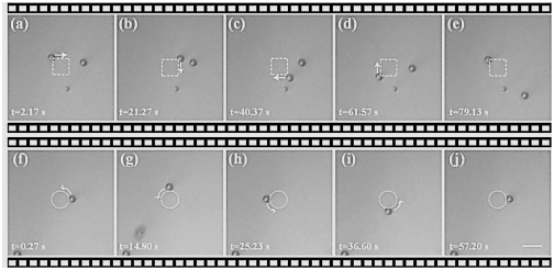
Fig.3.Experimental Results that Laser Captures and Manipulates Particles after Penetrating the Scattering Medium. (a)-(e)Manipulating particles to move along the rectangular locus after scattering medium; (f)-(j)Manipulating particles to move along the circular locus after scattering medium (Gauge: 10μm)
In the third paper “Three-dimensional Space Optimization for Near-field Ptychography”, 3D space optimization for near-field ptychography is realized. Ptychography is a lens-free coherent diffractive imaging technology and boasts advantages of large view field, high resolution and quantitative phase. By recording multi-piece crossover diffraction image, the use of crossover-area data redundancy and advanced phase retrieval algorithm can retrieve object’s transmittance function distribution, resolution coherent state and calibration system error. This lens-free imaging method has been successfully applied in visible light, electron wave band and X-ray wave band. In actual application, however, ptychography is still subject to certain limitations. For example, in imaging of 3D thick sample, the thickness is unknown. Conventional imaging methods work to reduce the imaging thickness of every layer of samples, which often leads to increase of imaging layers. Moreover, the method is only suitable for continuous samples, and may cause pseudo image of the samples scattered and with uneven space distribution. Additional blank layers may also lower quality of image.
This paper proposes a genetic algorithm-based 3D ptychographical imaging algorithm (GA-3ePIE), which can optimize layer number and layer distance simultaneously, and is suitable for near-field 3D Ptychography. Compared to far field, it can restructure the view field of the same size with less images, and raise lower requirements for light source coherence and the dynamic range of the probe. The analysis reveals that with the crossover rate and sampling rate higher, the layers retrievable become more. The algorithm can also be popularized to X-ray and electron wave band field, and be used in other computational imaging technology, such as Fourier ptychographical microscopic imaging technology.
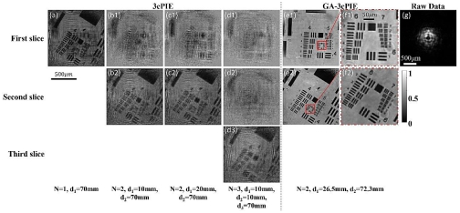
Fig.4. Intensity Retrieval Results of USAF Resolution Board under Different Parameters. (a) Sing-layer restructuring results. (b1-b2) and (c1-c2) Two-layer restructuring results with different layer distance. (d1-d3) Three-layer restructuring results, including a blank layer. (e1-e2) and (f1-f2) Restructuring results with GA-3ePIE algorithm and the enlarged image. (g) A typical diffraction image.
Worth mentioning, the article “Single-shot Slightly Off-axis Digital Holographic Microscopy with Add-on Module based on Beamsplitter Cube” in the same issue of Optics Express by a Spanish scholar cited 7 related papers by our research group, which is an affirmation of our works in digital holographic microscopic imaging field and highlights our contributions and influences in the field.
The team headed by Research Fellow Yao Baoli has been working on the research and development of new methods, technologies and instruments for new-type optical imaging and optical micro manipulation, and has published more than 200 papers in famous international journals such as PRL, PRA, OL and OE, and been granted multiple national invention patents. The team has won the first and second awards of Shaanxi Province for science and technology and the honorary titles such as Key Sci-tech Innovation Team of Shaanxi Province. In 2013, it firstly proposed and realized the digital micro-mirror device (DMD) and LED lighting-based microscopic imaging technology of structured light illumination, with the resolution up to 90nm. Relevant imaging equipment has been successfully applied to multiple life science studies. The team has researched and developed multiple sets of laser optical tweezers micro manipulation instruments for many universities both at home and abroad, which have demonstrated stable and reliable performance and won good remarks from users generally.
The Nobel Prize in Chemistry 2014 was awarded for ultra-resolution microscopic imaging technology and the Nobel Prize in Physics 2018 for laser optical tweezers and CPA technology, which the National Key Laboratory for Transient Optics and Photon Technology of our institute met by chance in terms of research orientation, strengthening our research confidence. Only by conducting basic research in a down-to-earth way, unremittingly and perseveringly and carrying out domestic and international cooperation and academic exchange actively, can new academic thought be generated and innovative works be created. (By the Key Laboratory)