Recently, the Super-resolution Imaging Innovation Team under the National Key Laboratory for Transient Optics and Photon Technology, XIOPM, through cooperation with Yang Xingke Team under the Institute of Zoology, Chinese Academy of Sciences, successfully applied the structured illumination microscopy developed by them to acquisition of 3D structures of small organisms in a fast high-resolution format, providing direct scientific research evidences for deep bio-morphological research of bio-evolution, classification (branch) and exotic species discrimination. The research achievement was published in the Frontiers in Zoology in June 2016, with Doctor Dan Dan from our institute as the lead author.
The know-how used in the research is the structured illumination microscopic technology based on DMD device modulation and LED lighting developed by our Yao Baoli and Lei Ming Team in 2013, or D-SIM for short (Scientific Reports 2013, Invention Patent No.: ZL201110448980.8). It uses high-speed digital microscopic device (DMD) and high-brightness LED source to generate structural light field, able to realize super-resolution imaging and be used for 3D tomography imaging. Compared with traditional 3D imaging technologies such as confocal laser scanning microscopy (CLSM), etc, D-SIM not only boasts higher spatial resolution, shorter imaging time (generally only needs decades of seconds to few minutes) and better imaging quality, but also greatly reduces device complex and cost. Under 3D tomography mode, the technology was designed mainly for observation of specimens of submillimeter magnitude, but met centimeter-magnitude even larger sizes of many bio-specimens in fact. In order to solve the problem, through concerted efforts, both teams proposed a solution specially addressing fast 3D imaging of small organisms. With invertebrates representing six different categories as the specimens, through comparison with currently popular 3D imaging technologies such as CLSM, Micro-CT, FIB-SEM and MRI, the teams found that D-SIM is superior to the existing technologies in terms of small-organisms fine-3D-structure imaging quality, imaging speed and specimen preparation method, and has great advantages over traditional optical microscopy in terms of compatibility and equipment cost. Therefore it is hopeful of becoming a common imaging unit in ordinary labs.
Actual application and solving problem in application are effective ways to test and embody value of self-developed science instruments. Successful application to bio-morphological research of the science instrument developed by XIOPM provides a new technical means for research in the field. The achievement not only embodies abilities of the research personnel in continuously tackling and innovating but shows that XIOPM has taken another solid step in multi-disciplinary development and research achievement translation. At present, both teams are carrying out deeper cooperation and are expected to make more high-level research achievements.
Article linkage: http://frontiersinzoology.biomedcentral.com/articles/10.1186/s12983-016-0158-9
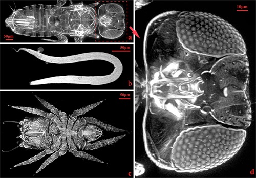
Small Organisms 3D Imaging Result 1 (a. Parasitoid wasp; b. Nematode; c. Mite, d. Partial enlargement of parasitoid wasp head)
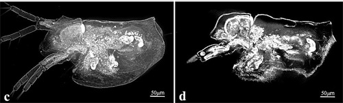
Small Organisms 3D Imaging Result 2 (c. Overall image of water flea; d. Sectional image of a part in water flea body)